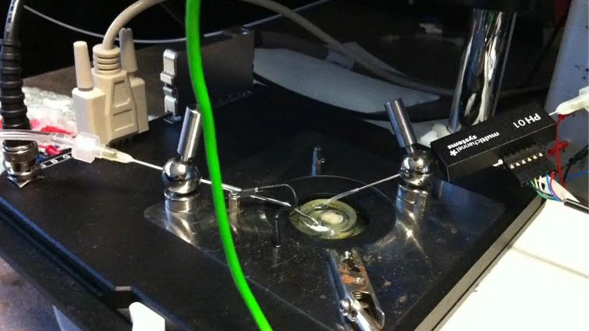
These interneurons are believed to act as “local brakes” mediating the flow of communication through these circuits. We are currently interested in how alterations in these neurons may underpin fronto-temporal dementia and Autistic spectrum disorder. Beyond GABAergic interneurons, we are also investigating how alterations in the connectivity of the basal ganglia, specifically the connections between striatal territories and pallidal territories may also contribute to these atypical behavioural states. In our lab we have all the necessary equipment to prepare and keep mouse brain slices viable (alive) exvivo. We also have advanced electrophysiological recording equipment to allow invitro extracellular neuronal recordings from acute slice preparations. The technique we employ is called a multi-electrode array (MEA). MEAs have sixty electrodes made from titanium nitride which are embedded in a glass slide. The electrodes are spaced 200µm apart and are 30µm in size. MEAs have excellent spatial and temporal precision so large amounts of data can be accurately collected in a relatively short space of time. MEAs also reduce the number of animals needed to achieve acceptable levels of power compared to other electrophysiological techniques, such as patch clamping. MEAs can be set up to allow for healthy slice preparations and recording for several hours (Clark, 2020; Clark & Bracci, 2018)). As a high throughput technique MEAs are well suited for investigating the effect of pharmacological manipulations on tonically active brain regions. As well as enabling extracellular recording of acute slice preparations the MEA can also be configured to stimulate brain regions / neurons. This enables our lab to investigate both tonic and evoked activity and dissect the circuitry of subcortical brain regions. As well as acute slice recording techniques we also have all the necessary equipment for visualisation of fixed slice preparations, including numerous Immunohistochemical techniques.

