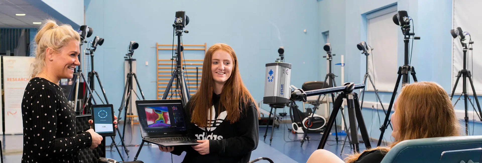
Thermal Imaging
Thermal imaging is possible at UCLan through a Flir A40 camera, from which data can be analysed in standard and custom written software. Thermal images may be quantified. The mean temperature of a specific area can be recorded, or a temperature distribution over a specific area. There are links between with the skin surface temperature and muscle function, however there are also clear links with the sympathetic nervous system. Sympathetic fibres are very thin and unmyelinated. They are probably therefore especially vulnerable to damage. When sympathetic fibres are disturbed changes in the vasomotor activity can be expected in the innervation area and changes in skin temperature follow (Comstok 1986). The exploration skin surface temperature may therefore provide insight into the function of the affected sympathetic fibres. Tchou (1992) stated that all vasoconstrictor nerves belong to the sympathetic system, therefore skin temperature is an indirect measure of sympathetic activity. As skin has an emissivity (Heat transfer coefficient) of 0.98, it is essentially transparent to heat and acts as a window to the vasculature beneath it.
Isokinetic Dynamometer - Cybex NORM
Isokinetic testing was originally a tool used mainly in exercise science and the only isokinetic movements available were concentric (with no thought given to isotonics, isometrics, continuous passive motion (CPM) or range of motion expansion). Eventually, isokinetics found its way into therapy albeit with very rigid machines usually specific to a joint or small numbers of joints. In the early 1980's (around 1984) the world of isokinetics changed dramatically. Servo motors and microprocessors transformed the early machines into fast and dynamic tools offering instant data analysis and reproducibility. This now meant that real time data became available and testing became as important as exercise. Today isokinetic dynamometers are used for the measurement maximal isometric contractions, muscle balance tests, pathology diagnosis and in rehabilitation, these machines are now incredibly versatile allowing for measurements from almost all of the joints of the body and with additional kits to test functional activities such as driving or opening a door.
Electromyography EMG
Electromyography (EMG) is the electrical activity associated with muscle activation. Nerve impulses cause twitch responses in muscle fibres, these combined cause the bulk contraction of a muscle. This is produced by the depolarizing of motor units, often referred to as Motor Unit Action Potential (MUAP). The electrical signal produced has a magnitude and a frequency response. Both these measurable features allow not just the onset of the muscle action but the size of the contraction in the form of motor unit recruitment but also the rate of depolarizing in the form of changes in the frequency response. This latter feature is particularly important in the study of fatigue.
Pressure Measurement
Excessive pressures can cause tissue damage. Pressure can be a main cause of ulceration. Assessment of pressure may be able to predict when and where ulceration might occur, allowing the clinician to prevent serve damage taking place.
The F-Scan ® system provides bipedal plantar pressure and force measurements on the feet using paper-thin reusable sensors placed in to the patients’ shoes. The system instantly detects, displays, and records plantar pressures and forces without interfering with normal gait pattern for the truest measurements available. The quantitative information shown will enhance your ability to evaluate, substantiate, and document your diagnosis. With the F-Scan system, you get a view from inside the shoe.
MVN BIOMECH Motion Capture Suit
The XSENS MVN motion capture suit uses miniature three-dimensional (3D) gyroscopes, accelerometers and magnetometers. This system requires no cameras and do not require markers to be ‘seen’. Therefore they can be used indoors and outdoors regardless of lighting conditions. The combination of the three types of sensor allows the tracking of each segment in six degrees of freedom of up to 17 body segments.
Near Infrared Oxygenation Monitor
Non-invasive, continuous measurement of tissue oxygen levels. The NIRO monitors tissue oxygen levels using near infrared spectroscopy. In addition to measuring the tissue oxygenation index (TOI), which indicates tissue oxygen saturation and the normalized tissue haemoglobin index (nTHI) which indicates blood concentration using a low intensity light that is safe for the body, changes in oxygenated, deoxygenated and total haemoglobin concentrations (ΔO2Hb, ΔHHb and ΔcHb) are measured in real time. The NIRO measures tissue oxygen levels using near infrared spectroscopy. A low intensity light that is safe for the body is used to measure tissue oxygen saturation (TOI) and blood concentration (nTHI) in real time. The greatest strengths are displayed in cases where management of soft tissue oxygenation is important. The particular focus of the use of this equipment is studies on muscle tissue oxygenation for sports medicine and rehabilitation.

